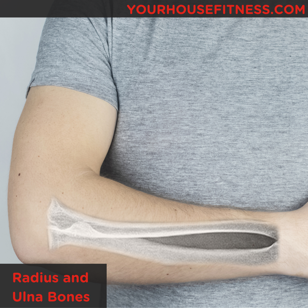
Bones of the Forearm: The Radius and the Ulna
Table of Contents
What Are the Radius and Ulna
The body is made up of hundreds of bones! It can be hard to keep track of them all. Two of the most important bones in the arm are the Radius and Ulna. These two long bones are located in the forearm and work together to produce movement at the wrist and elbow.
An easy way to remember which bone is on each side of the forearm is the saying, ‘the Radius Rotates’. When you turn your palm to face the floor, the Radius crosses over the Ulna. This is an easy way to remember that the radius is located on the lateral (or outside) of the forearm and the Ulna is located on the medial (or inside) of the forearm.
Ulna and Radius Diagram
What Is the Function of the Radius and Ulna
The primary function of the Radius is to work with the Ulna at the elbow to produce pronation and supination. This allows us to rotate our palms towards the ceiling and down towards the floor.
The primary function of the Ulna is to articulate with the Humerus to form the elbow joint. As well, the Ulna creates the Radio-ulnar joint with the Radius at the wrist to produce pronation and supination of the wrist.
Muscles That Are Attached to the Radius and Ulna
There are many muscles in the forearm that attach to the Radius and the Ulna.
Muscles Attach to the Radius
Muscles Attach to the Ulna
Flexor Carpi Ulnaris
Flexor Digitorum Profundus
Flexor Digitorum Superficialis
Pronator Quadratus
Supinator
Abductor Pollicus Longus (originates from interosseous membrane)
Extensor Pollicus Longus (originates from interosseous membrane)
Extensor Indicis (originates from interosseous membrane)
Radius and Ulna Fracture
Both the Radius and the Ulna are susceptible to fractures. Fractures to the Radius and the Ulna can occur at the wrist, in the middle (or shaft) of each bone, and proximally near the elbow. There are many ways to fracture these bones. The most common causes of fracture are falling on an outstretched arm, a direct blow to the arm, or a motor vehicle accident. Symptoms of a Radius and/or Ulna fracture include swelling, bruising, pain, numbness, and the inability to move the arm.
You will need to see a physician if you suspect that you have fractured your forearm. They will perform an x-ray to confirm if you have indeed suffered a fracture, the type of fracture, and the area the fracture is located. Forearm fractures can require surgery so that the bones heal correctly. It is critical that the Radius and Ulna are able to stabilize each other, so they need to be set up to heal correctly. If you do not require surgery, a reduction can be done to move the bones into place before a splint is placed on the forearm. Medication can be used to help reduce pain.
It is important to remember that there are different types of fractures and as a result different types of treatments, procedures, and complications. This is why it is very important to see a healthcare professional if you believe that you have fractured the Radius and/or the Ulna to ensure that your recovery process is initiated correctly.
Radius and Ulna Fracture Complications
As with any injury, there are different fracture complications that can arise. Depending on the type of fracture, sharp pieces of the bone fragment can tear the surrounding tissues and damage blood vessels. If you have suffered from an open fracture, the bone will be visible and exposed to the environment outside of the body. This creates the opportunity for the bone to be contaminated and infected. Infections of the bone are extremely serious and can require multiple surgeries and medications to recover.
One of the most significant Radius and Ulna fracture complications is acute compartment syndrome. Acute compartment syndrome occurs when there is excessive swelling in the forearm. This excessive swelling can cut off the blood supply to the arm and result in significant pain when attempting to move the fingers. To relieve this pressure, the forearm is surgically opened so that the pressure can be released, and the blood flow returned to normal.
Ulna and Radius Labeled
A labeled image of the Ulna and Radius is a useful tool to visually understand where the landmarks are on each bone.
Difference Between Radius and Ulna
Although the Radius and the Ulna appear to be very similar at a first glance, they do have their differences. For instance, the Radius is thicker than the Ulna, but the Ulna is slightly longer than the Radius.
The Radius also runs along the lateral side of the forearm, from the elbow to the thumb and the Ulna runs along the medial side of the forearm to the pinky finger. Lastly, The Radius and the Ulna also have different muscles that attach to each bone and different landmarks on each bone.
Radius and Ulna Bone Markings
The Radius and the Ulna both have landmarks that are important for different muscle and joint attachments.
Bone Markings on the Radius
Head of the Radius
Styloid Process
Ulnar notch
Bone Markings on the Ulna
The radial notch of the Ulna
Olecranon process
Trochlear notch
Coronoid process
Styloid process


